Caesarean
section
- Local anaesthesia is a safe alternative to general, ketamine or
spinal anaesthesia when these anaesthetics or persons trained in their
use are not available;
- The use of local anaesthesia for caesarean section requires that
the provider counsel the woman and reassure her throughout the
procedure. The provider should use instruments and handle tissue as
gently as possible, keeping in mind that the woman is awake and alert.
Note: In the case of heart failure, use local infiltration
anaesthesia with conscious sedation. Avoid spinal anaesthesia.
- an inaccessible lower segment due to dense adhesions from previous
caesarean sections;
- transverse lie (with baby’s back down) for which a lower uterine
segment incision cannot be safely performed;
- fetal malformations (e.g. conjoined twins);
- large fibroids over the lower segment;
- a highly vascular lower segment due to placenta praevia;
- carcinoma of the cervix.
If the
baby’s head is deep down into the pelvis as in obstructed
labour,
prepare the vagina
for assisted caesarean delivery.
Have the operating table tilted to the left or place a pillow or
folded linen under the woman’s right lower back to decrease supine
hypotension syndrome.
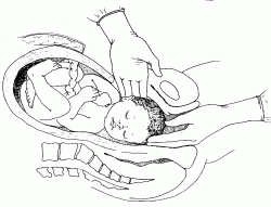
OPENING THE ABDOMEN
Note: If the
caesarean section is peformed under local anaesthesia, make the
midline incision that is about 4 cm longer than when general anaesthesia
is used. A
Pfannenstiel incision should not be used as it takes longer,
retraction is poorer and it requires more local anaesthetic.
Figure P-19
Site of abdominal incision

-
Hold the fascial edge with forceps and lengthen the incision up
and down using scissors.
Use fingers or scissors to separate the rectus muscles (abdominal
wall muscles).
Use fingers to make an opening in the peritoneum near the
umbilicus. Use scissors to lengthen the incision up and down in order to
see the entire uterus. Carefully, to prevent bladder injury, use
scissors to separate layers and open the lower part of the
peritoneum.
Place a bladder retractor over the pubic bone.
Use forceps to pick up the loose peritoneum covering the anterior
surface of the lower uterine segment and incise with scissors.
Extend the incision by placing the scissors between the uterus
and the loose serosa and cutting about 3 cm on each side in a transverse
fashion.
Use two fingers to push the bladder downwards off of the lower
uterine segment. Replace the bladder retractor over the pubic bone and
bladder.
OPENING THE UTERUS
• Use a scalpel to make a 3 cm transverse incision in the lower
segment of the uterus. It should be about 1 cm below the level where the
vesico-uterine serosa was incised to bring the bladder down.
• Widen the incision by placing a finger at each edge and gently
pulling upwards and laterally at the same time (Fig P-20).
• If the
lower uterine segment is thick and narrow, extend the incision
in a crescent shape, using scissors instead of fingers to avoid
extension of the uterine vessels.
It is important to make the
uterine incision big enough to deliver the head and body of the baby
without tearing the incision.
Figure P-20
Enlarging the uterine incision

DELIVERY OF THE BABY AND PLACENTA
To deliver the baby, place one hand inside the uterine cavity
between the uterus and the baby’s head.
With the fingers, grasp and flex the head.
Gently lift the baby’s head through the incision (Fig P-21),
taking care not to extend the incision down towards the cervix.
With the other hand, gently press on the abdomen over the top of
the uterus to help deliver the head.
If the
baby’s head is deep down in the pelvis or vagina, ask an
assistant (wearing high-level disinfected gloves) to reach into the
vagina and push the baby’s head up through the vagina. Then lift and
deliver the head (Fig P-22).
Figure P-21
Delivering the baby’s head

Figure P-22
Delivering the deeply engaged head
- ampicillin 2 g IV;
- OR cefazolin 1 g IV.
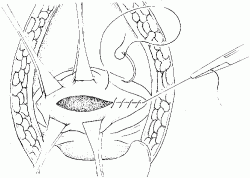
CLOSING THE UTERINE INCISION
Note: If a Couvelaire uterus (swollen and discolored by
blood) is seen at caesarean section, close it in the normal manner and
observe.
Grasp the corners of the uterine incision with clamps.
Grasp the bottom edge of the incision with clamps. Make sure it
is separate from the bladder.
Look carefully for any extensions of the uterine incision.
Repair the incision and any extensions with a continuous locking
stitch of 0 chromic catgut (or polyglycolic) suture (Fig P-23).
If there is any
further bleeding from the incision site, close with
figure-of-eight sutures. There is no need for a routine second layer of
sutures in the uterine incision.
Figure P-23
Closing the uterine incision
CLOSING THE ABDOMEN
Note: There is no need to close the bladder peritoneum or the
abdominal peritoneum.
If
there are signs of infection, pack the subcutaneous tissue with
gauze and place loose 0 catgut (or polyglycolic) sutures. Close the skin
with a delayed closure after the infection has cleared.
If there are
no signs of infection, close the skin with vertical mattress
sutures of 3-0 nylon (or silk) and apply a sterile dressing.
Gently push on the abdomen over the uterus to remove clots from
the uterus and vagina.
PROBLEMS DURING SURGERY
BLEEDING IS NOT CONTROLLED
BABY IS BREECH
- Deliver the legs and the body up to the shoulders, then deliver the
arms;
- Flex (bend) the head using the Mauriceau Smellie Veit manoeuvre.
BABY IS TRANSVERSE
THE BABY’S BACK IS UP
If the back is up (near the top of the uterus), reach into
the uterus and find the baby’s ankles.
Grasp the ankles and pull gently through the incision to deliver
the legs and complete the
delivery as for a breech baby.
THE BABY’S BACK IS DOWN
PLACENTA PRAEVIA
If a low anterior placenta is encountered, incise through
it and deliver the fetus.
After delivery of the baby, if the
placenta cannot be detached manually, the diagnosis is placenta
accreta, a common finding at the site of a previous caesarean scar.
Perform a
hysterectomy.
Women with placenta praevia are at high risk of postpartum
haemorrhage. If there is
bleeding at the placental site, under-run the bleeding sites
with chromic catgut (or polyglycolic) sutures.
Watch for bleeding in the immediate postpartum period and take appropriate action.
POST-PROCEDURE CARE
- Massage the uterus to expel blood and blood clots. Presence of
blood clots will inhibit effective uterine contractions;
- Give oxytocin 20 units in 1 L IV fluids (normal saline or Ringer’s
lactate) at 60 drops per minute and ergometrine 0.2 mg IM and
prostaglandins (Table
S-8). These drugs can be given together or sequentially.
- ampicillin 2 g IV every 6 hours;
- PLUS gentamicin 5 mg/kg body weight IV every 24 hours;
- PLUS metronidazole 500 mg IV every 8 hours.
HIGH VERTICAL (“CLASSICAL”) INCISION
- Check the position of the round ligaments and ensure that the
incision is in the midline (the uterus may have twisted to one
side);
- Make the uterine incision in the midline over the fundus of the
uterus;
- The incision should be approximately 12-15 cm in length and the
lower limit should not extend to the utero-vesical fold of the
peritoneum.
Ask an assistant (wearing high-level disinfected gloves) to apply
pressure on the cut edges to control the bleeding.
Cut down to the level of the membranes and then extend the
incision using scissors.
After rupturing the membranes, grasp the baby’s foot and
deliver the baby.
Deliver the placenta and membranes.
Grasp the edges of the incision with Allis or Green Armytage
forceps.
Close the incision using at least three layers of suture:
- Close the first layer closest to the cavity but avoiding the
decidua with a continuous 0 chromic catgut (or polyglycolic) suture;
- Close the second layer of uterine muscle using interrupted 1
chromic catgut (or polyglycolic) sutures;
- Close the superficial fibres and the serosa using a continuous 0
chromic catgut (or polyglycolic) suture with an atraumatic needle.
The woman should not labour with
future pregnancies.
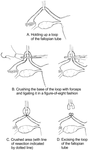
TUBAL LIGATION AT CAESAREAN
Tubal ligation can be done immediately following caesarean section if
the woman requested the procedure
before labour began (during prenatal visits). Adequate
counselling and informed decision-making and consent must precede
voluntary sterilization procedures; this is often not possible during
labour and delivery.
Review for consent of patient.
Grasp the least vascular, middle portion of the fallopian tube
with a Babcock or Allis forceps.
Hold up a loop of tube 2.5 cm in length (Fig P-24 A).
Crush the base of the loop with artery forceps and ligate it with
0 plain catgut suture (Fig P-24 B).
Excise the loop (a segment 1 cm in length) through the crushed
area (Fig P-24).
Repeat the procedure on the other side.
Figure P-24
Tubal ligation
Top of page


